


PHINDER STUDY RESULTS
Establishing a path to PH detection
The preliminary results from the PHINDER study show the opportunity of using routine ILD tests often to screen for PH.2
Background & methodology
The PHINDER study is a multicenter, noninterventional, prospective study that aims to identify parameters associated with the presence of PH in ILD and develop a PH detection tool to apply in practice (N=300-350).2
This preliminary analysis is presented to inform current practice, while the final dataset will be used to establish prognostic thresholds and validate a structured screening algorithm.
Inclusion criteria: To enroll, patients must meet ≥1 criterion from ≥2 different enrichment categories based on assessments within 180 days of screening.2*

Noninvasive assessments
An enriched population of patients underwent a broad range of assessments.2†


Questionnaire
Investigators completed the Investigator’s Suspicion of PH Questionnaire.2

RHC
Patients underwent RHC to confirm precapillary PH.2‡
Enrichment categories2
- 1. PFTs and/or HRCT
- 2. Signs and symptoms
- 3. Echocardiography
This study is preliminary data that should be reviewed with appropriate caution.
Preliminary findings
DLCO decrease and pulmonary artery enlargement on HRCT may be predictive of PH-ILD2§

PFT KEY SIGNALS2:
- Decreased DLCO
- Elevated FVC/DLCO ratio

HRCT KEY SIGNALS2:
- Elevated PA/aorta ratio
- Elevated RV/LV ratio
Implications: Routine ILD tests can help predict PH.
Compared to RHC, gestalt-based assessments may be inaccurate2
In the PHINDER study, preliminary results showed that RHC confirmed PH in one-third of patients labeled as having a "low likelihood" of PH.2

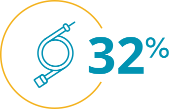
of patients labeled "low likelihood" to have PH were confirmed to have PH via RHC.2
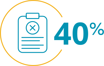
of gestalt-based assessments were inaccurate compared to RHC.2
Additional study limitations
- The study analyses are ongoing, which may introduce bias or limit the robustness of preliminary findings
- Lack of contrast may have reduced the accuracy of vascular measurements, especially pulmonary artery evaluation
- The statistical analyses may also be limited by the sample size of subgroups and the fact that outliers were not removed
- This analysis did not examine differences by sex; future studies with adequately powered male and female groups are needed to explore potential sex-based differences
- A univariate model was used for this preliminary data; a more robust multivariate analysis will be performed on the final dataset
- The time gap between noninvasive tests and RHC could have affected results due to potential disease progression, although the median interval was short
- The study described in these reprints was sponsored, designed, and executed by United Therapeutics
*PH-enrichment categories are: 1. PFTs and/or HRCT; 2. Exercise capacity, oxygenation, biomarkers, and/or physical exam; 3. Echocardiography.2
†Assessments included PFTs, HRCT, exercise capacity, biomarkers, physical exam features, and echocardiography.2
‡Precapillary PH defined as mPAP >20 mm Hg, PAWP ≤15 mm Hg, and PVR >2 WU.2
§HRCT and echocardiography imaging were centrally reviewed and interpreted but did not account for inter- or intra-reader variability. The absence of contrast-enhanced HRCT may have limited the precision of vascular measurements.2
‖Following all noninvasive assessments, investigators were asked to rank their suspicion of PH in each patient as high, medium, or low.2
¶One patient did not have Investigator's Suspicion of PH reported prior to RHC.2
DLCO=diffusing capacity of the lung for carbon monoxide; FVC=forced vital capacity; HRCT=high-resolution computed tomography; ILD=interstitial lung disease; LV=left ventricular; mPAP=mean pulmonary arterial pressure; PA=pulmonary artery; PAWP=pulmonary arterial wedge pressure; PFT=pulmonary function test; PH=pulmonary hypertension; PVR=pulmonary vascular resistance; RHC=right heart catheterization; RV=right ventricular; WU=Wood units.
The information contained in this section of the site is clinical in nature and specifically created for healthcare professionals. If you are not a US healthcare professional, please click CLOSE to return to the consumer section of the site.